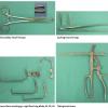
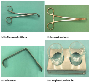
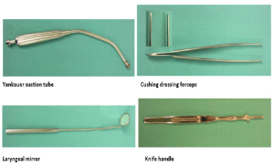
Dr. Richard Smith preferred method:
- Informed consent was reviewed with the parents. The patient was transferred to the room and a time out was performed. A Crowe-Davis mouth gag was placed with good exposure of the oropharynx. First the adenoid bed was addressed. The palate was palpated and uvula inspected with no abnormalities noted. Sizing the adenoid curette on the maxillary incisors, the curette was placed at the base of the nasopharynx and the adenoids were removed. A tonsil ball was used to pack the adenoid bed. The right tonsil was then addressed. Curved Allis clamps were placed on the superior pole, and the tonsil was medialized. A Cushing toothed forceps was used to perform blunt dissection of the superior pole of the tonsil, being careful not to enter the tonsil, but to dissect out the fibrous capsule. Once a significant portion of the tonsil was free, with only a portion of the inferior pole remaining, a Tyding tonsil snare was placed around the remaining tonsil tissue, and the tonsil was subsequently removed. Two tonsil balls were placed in the right tonsillar fossa. The left tonsil was then addressed, and removed in a similar fashion as the right, with subsequent hemostasis achieved using suction cautery. The tonsil balls were then removed from the tonsillar and adenoidal beds with similar hemostasis achieved using suction cautery. 6.0 cc of Marcaine was injected along the bilateral palatoglossal and palatopharyngeal arches. The nasal and oral cavities were thoroughly irrigated with no active bleeding noted. The Crowe-Davis retractor was removed at this time. Irrigation was performed and the patient's stomach was suctioned for contents. The mouth was cleaned of debris and wiped clean, and bacitracin ointment was placed on the lips. The patient was then turned over to Anesthesia in good condition.
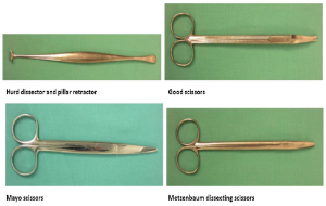
- Informed consent was reviewed with the parents and the patient was brought to the room and placed in the supine position. A Crowe-Davis retractor was placed with good visualization of the oropharynx. A red rubber catheter was then placed to retract the soft palate and allow good visualization. The soft palate was palpated, and uvula inspected with no abnormalities. An Adenoid curette was sized using the maxillary incisors and used to remove the adenoid bed. A tonsil ball was placed to help achieve hemostasis. The right tonsil was then addressed. Curved Allis clamps were used to grasp the superior pole of the tonsil and medialize it. The mucosa from the superior pole enveloping the fibrous capsule was divided using a sickle knife. Good scissors were used to dissect the tonsil from the tonsillar bed, maintaining the fibrous capsule. With only the inferior pole remaining, a Tydings tonsil snare was placed and the remaining tonsil was removed. A tonsil ball was placed into the tonsillar fossa for hemostasis. The left tonsil was then addressed. Curved Allis clamps were used to grasp the superior pole of the tonsil and medialize it. The mucosa from the superior pole enveloping the fibrous capsule was divided using a sickle knife. Good scissors were used to dissect the tonsil from the tonsillar bed, maintaining the fibrous capsule. With only the inferior pole remaining, a Tydings tonsil snare was placed and the remaining tonsil was removed. A tonsil ball was placed into the tonsillar fossa for hemostasis. The tonsil balls were removed, and suction cautery was used to achieve hemostasis in the tonsillar and adenoid beds. The nasopharynx was irrigated and suctioned, and the red rubber catheter was removed. Marcaine was injected into the tonsillar arches, and the Crowe-Davis was removed. The patients mouth was wiped clean, and ointment was placed prior to returning the patient to Anesthesia in good condition.
- Informed consent was reviewed with the patient and family immediately preoperatively after which the patient was brought to the room and placed in the supine position. A Crowe-Davis retractor was placed with good visualization of the oropharynx. The soft palate was palpated, and uvula inspected with no abnormalities. With a curved tonsil injection needle 1% lidocaine with 1:100,000 epinephrine was injected into the tonsillar pillars and deep to the tonsil. A waiting period (to permit maximal vasoconstrictive effect) was followed by addressing the right tonsil by grasping it with a curved allis clamp. The mucosa from the superior pole enveloping the fibrous capsule was divided using a sickle knife. A tonsillar hemostat (or Kelley) was used to develop a plane between the tonsil and fascia overlying the constrictor muscle. Individual blood vessels are identified and addressed using the guarded bipolary cautery. The tonsil scissors are used to incise mucosa and with a pushing action, (using scissors, Fischer knife, hemostat or bipolar cautery) the tonsil was delivered inferiorly to the lower pole. Bipolar cautery at the lower pole then permitted final separation of the tonsil from the fossa with scissors in a bloodless fashion. The tonsillar bed was carefully inspected with additional hemostasis accomplished with bipolar cautery supplemented (rarely) with 3-0 chromic suture placed with needle driver. Tannic acid is then placed in the fossa. An identical procedure is performed on the contralateral side and the procedure terminated. Marcaine may be injected into the tonsillar arches.
- Informed consent was obtained and the patient was transferred to the ASC and placed in the supine position. A Crowe-Davis retractor was placed with good visualization of the oropharynx. A curved Allis was used to grasp the superior pole of the right tonsil. Monopolar cautery was used to release the thin layer of mucosa from the fibrous layer of the tonsil. The tonsil was dissected out of the tonsillar bed with monopolar cautery while maintaining the fibrous layer of the tonsil. Two tonsil balls were placed for hemostasis of the tonsillar bed. The left tonsil was then addressed. A curved Allis was used to grasp the superior pole of this tonsil as well. Monopolar cautery was used to release the thin layer of mucosa from the fibrous layer of the tonsil. The tonsil was dissected out of the tonsillar bed with monopolar cautery along the fibrous capsule as well. Two tonsil balls were placed in this side as well. The tonsil balls were subsequently removed from both sides at that point, and suction cautery was used to achieve hemostasis of the tonsillar beds. The nasopharynx and oropharynx were copiously irrigated and suctioned. Marcaine was injected at 6 cc along the tonsillar pillars. The stomach was suctioned, and the patient was returned to anesthesia in good condition.
- Informed consent was reviewed, and the patient was brought to the OR and placed in the supine position. The patient underwent general oral endotracheal intubation. The table was rotated, patient positioned, eyes taped, and head was draped. A Crowe-Davis retractor was placed with good visualization of the oropharynx. A red rubber catheter was then placed to retract the soft palate and allow good visualization. The soft palate was palpated, and uvula inspected with no abnormalities. The tonsils were resected in the capsular plane using blunt dissection and coblation. The coblator tip cautery was used as needed. The nasopharynx and hypopharynx were irrigated and suctioned. Hemostasis was confirmed. The red rubber catheter and Crowe-Davis retractor were removed. There was no injury to the lips gums, teeth or tongue. The patient was awakened, extubated, and taken to recovery.
- Informed consent was reviewed with the parents and the patient was brought to the room and placed in the supine position. A Crowe-Davis retractor was placed with good visualization of the oropharynx. A red rubber catheter was then placed to retract the soft palate and allow good visualization. The soft palate was palpated, and uvula inspected with no abnormalities of signs of submucous cleft. An adenoid curette was sized using the maxillary incisors and used to remove the adenoid bed. A tonsil ball was placed to help achieve hemostasis. The right tonsil was then addressed. Curved Allis clamps were used to grasp the superior pole of the tonsil and medialize it. The mucosa from the superior pole enveloping the fibrous capsule was divided using a 14-blade scalpel. A Fischer knife was used to dissect the tonsil from the tonsillar bed, maintaining the fibrous capsule. With only the inferior pole remaining, a Tydings tonsil snare was placed and the remaining tonsil was removed. A tonsil ball was placed into the tonsillar fossa for hemostasis. The left tonsil was then addressed. Curved Allis clamps were used to grasp the superior pole of the tonsil and medialize it. The mucosa from the superior pole enveloping the fibrous capsule was divided using a 14-blade scalpel. A Fischer knife was used to dissect the tonsil from the tonsillar bed, maintaining the fibrous capsule. With only the inferior pole remaining, a Tydings tonsil snare was placed and the remaining tonsil was removed. A tonsil ball was placed into the tonsillar fossa for hemostasis. The tonsil balls were removed, and suction cautery was used to achieve hemostasis in the tonsillar and adenoid beds. The nasopharynx was irrigated and suctioned, and the red rubber catheter was removed. Marcaine was injected into the tonsillar arches, and the Crowe-Davis was removed. The patients mouth was wiped clean, and ointment was placed prior to returning the patient to Anesthesia in good condition.
REFERENCES
Erickson BK, Larson DR, St Sauver JL, Meverden RA, Orvidas LJ. Changes in incidence and indications of tonsillectomy and adenotonsillectomy, 1970-2005. Otolaryngol Head Neck Surg. 2009 Jun;140(6):894-901.
Toh A, Mullin A, Grainger J, Uppal H. Indications for tonsillectomy: are we documenting them? Ann R Coll Surg Engl. 2009 Nov;91(8):697-9.
Sia KJ, Tang IP, Kong CK, Nasriah A. Grisel's syndrome: a rare complication of tonsillectomy.. J Laryngol Otol. 2012 Feb 24:1-3.
Berry S, Pascal I, Whittet HB: Tonsillectomy a chaud for quinsy: revisited. Eur Arch Otorhinolaryngol. 2008 Jan;265(1):31-3
Akin RC, Holst R, and Schousboe LP. Rick factors for post-tonsillectomy haemorrhage. Acta otolaryngol. 2012 Jul:132(7):773-7

